
When faced with complex dental injuries, many patients in London search online to understand the diagnostic process and what advanced imaging might reveal about their condition. Dental trauma can affect multiple structures simultaneously, making accurate assessment crucial for appropriate treatment planning.
CBCT (Cone Beam Computed Tomography) 3D imaging has revolutionised how dental professionals evaluate complex injuries, particularly those involving facial trauma, root fractures, or jaw-related concerns. This advanced diagnostic technology provides detailed three-dimensional views that traditional X-rays cannot capture, helping clinicians understand the full extent of damage to teeth, bone, and surrounding tissues.
Understanding how CBCT 3D imaging works and when it may be recommended can help patients feel more informed about their dental care journey. This article explores the role of this advanced diagnostic tool in complex injury assessment, explaining how it supports clinical decision-making and contributes to comprehensive treatment planning. Professional dental evaluation remains essential for determining whether advanced imaging is appropriate for individual circumstances.
Experiencing these symptoms?
Delaying treatment can lead to tooth loss. We have slots available today.
What is CBCT 3D Imaging in Complex Dental Injuries?
CBCT 3D imaging provides detailed three-dimensional views of dental and facial structures, allowing clinicians to assess complex injuries with greater precision than traditional two-dimensional X-rays. This technology captures comprehensive images of teeth, bone, soft tissues, and their spatial relationships in a single scan.
How CBCT Technology Works in Dental Diagnostics
CBCT scanners rotate around the patient's head, capturing multiple images from different angles within seconds. Advanced computer software then reconstructs these images into detailed three-dimensional models that can be viewed from any direction. This technology operates using lower radiation doses compared to traditional medical CT scans whilst providing comprehensive, high-resolution detail of dental and maxillofacial structures.
The scanning process is typically quick and comfortable, with patients remaining seated whilst the equipment rotates around their head. The resulting images allow dental professionals to examine root structures, bone density, fracture patterns, and tissue relationships that may not be visible on conventional radiographs. This comprehensive view supports more accurate diagnosis and treatment planning for complex cases.
Meet Dr. Yasha Shirazi
Principal Dentist at Emergency Dentist London
"We treat hundreds of dental emergencies every month. The sooner you come in, the easier the fix usually is."
Book an appointment with our team →Clinical Applications in Complex Injury Assessment
CBCT 3D imaging proves particularly valuable when assessing multi-layered dental trauma. Complex injuries often involve damage to multiple tooth structures, surrounding bone, or adjacent teeth simultaneously. Traditional imaging may not reveal the full extent of such injuries, potentially leading to incomplete treatment planning.
This technology excels in evaluating root fractures, especially vertical fractures that may not appear clearly on standard X-rays. It can also assess the relationship between damaged teeth and important anatomical structures such as nerves, sinuses, or adjacent tooth roots — a consideration particularly relevant when evaluating the proximity of wisdom teeth to nerve pathways. For jaw-related injuries, CBCT imaging helps evaluate bone fractures, joint positioning, and the impact on surrounding structures.
The three-dimensional perspective allows clinicians to measure distances accurately, assess bone quality, and identify complications that might affect treatment outcomes. This detailed information supports more precise treatment approaches and helps patients understand their condition more thoroughly.
Understanding Complex Dental Injury Patterns
Complex dental injuries rarely affect just one structure. A single traumatic event might damage tooth enamel, dentine, pulp tissue, periodontal ligaments, and surrounding bone simultaneously. Understanding these interconnected injury patterns helps explain why comprehensive imaging becomes important for thorough assessment.
Root fractures may extend in multiple directions, creating fracture lines that appear differently when viewed from various angles. Bone fractures might affect tooth stability, healing potential, or future treatment options. CBCT imaging reveals these relationships clearly, providing the detailed information needed for comprehensive treatment planning.
The technology also helps identify injuries that might not cause immediate symptoms but could lead to complications later. Early detection of such issues supports proactive treatment approaches and may help prevent more extensive problems developing over time.
When CBCT 3D Imaging May Be Recommended
Dental professionals may consider CBCT imaging when conventional radiographs provide insufficient information for accurate diagnosis or treatment planning. This typically occurs with complex trauma cases, suspected root fractures, or when multiple treatment options are being evaluated.
Symptoms that might indicate the need for advanced imaging include persistent pain without clear cause on standard X-rays, unusual swelling patterns, or trauma involving multiple teeth or facial structures. However, the decision to recommend CBCT imaging always depends on individual clinical circumstances and professional judgement.
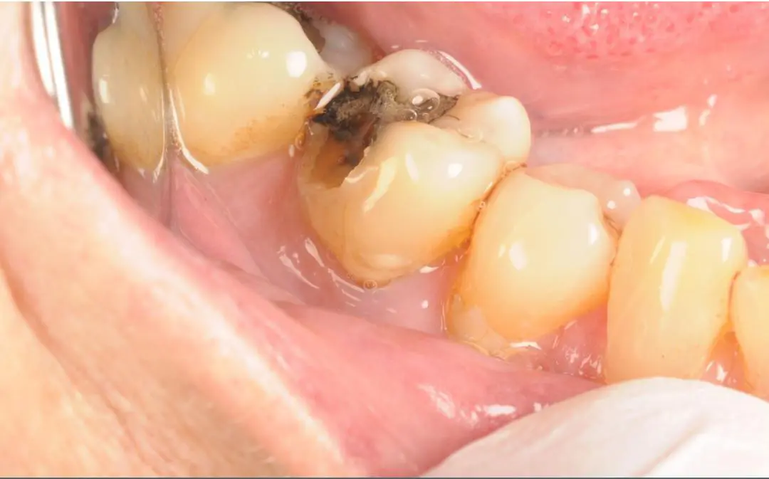
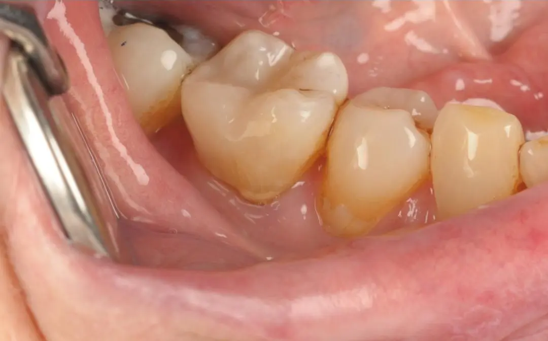
Real Patient Result: Emergency White Filling
Treatment by Dr Kamran
Patients experiencing facial trauma, suspected jaw fractures, or complex tooth damage may benefit from this detailed imaging approach. The three-dimensional view helps ensure that all aspects of the injury are identified and addressed appropriately during treatment planning.
Treatment Planning Benefits of 3D Imaging
Advanced imaging supports more precise treatment planning by providing comprehensive information about injury extent and complexity. This detailed view helps dental professionals explain treatment options more clearly and set realistic expectations about outcomes and healing timeframes.
The technology allows for virtual treatment planning, where different approaches can be evaluated before beginning actual treatment. This may include assessing the feasibility of tooth preservation procedures, planning surgical approaches, or determining the best sequence for multi-stage treatments.
For complex cases requiring specialist input, CBCT images provide valuable information for referral discussions and collaborative treatment planning. The detailed three-dimensional data supports communication between different dental professionals involved in comprehensive care.
Prevention and Oral Health Considerations
Whilst CBCT imaging primarily serves diagnostic purposes, understanding injury prevention remains important for long-term oral health. Wearing appropriate protective equipment during sports, avoiding hard foods that might damage teeth, and maintaining regular dental check-ups all contribute to injury prevention.
Good oral hygiene supports healing after any dental trauma, as healthy surrounding tissues typically recover more effectively than compromised ones. Regular professional assessments help identify and address minor issues before they develop into complex problems requiring advanced diagnostic procedures.
Understanding the signs of dental trauma and seeking prompt professional evaluation can help ensure that appropriate diagnostic methods, including advanced imaging when necessary, are used effectively to support optimal treatment outcomes.
Key Points to Remember
• CBCT 3D imaging provides detailed three-dimensional views that support comprehensive assessment of complex dental injuries
• This technology reveals injury patterns and structural relationships that may not be visible on conventional X-rays
• Professional evaluation determines when advanced imaging is appropriate for individual circumstances
• The detailed information supports more precise treatment planning and helps set realistic expectations
• Early professional assessment of dental trauma helps ensure appropriate diagnostic approaches are used
• Prevention through protective measures and regular dental care remains the best approach to avoiding complex injuries
Frequently Asked Questions
Is CBCT imaging safe for dental diagnosis?
CBCT scanners are designed specifically for dental and maxillofacial imaging, using lower radiation doses than traditional medical CT scans whilst providing detailed images. Dental professionals carefully consider the benefits and risks for each individual case, recommending advanced imaging only when the diagnostic information is essential for appropriate treatment planning.
How long does a CBCT scan take?
Most CBCT scans are completed within 10-40 seconds of actual scanning time, though the entire appointment may take 15-30 minutes including positioning and image processing. The procedure is typically comfortable, with patients remaining seated whilst the equipment rotates around their head. No special preparation is usually required.
What can CBCT imaging detect that regular X-rays cannot?
CBCT provides three-dimensional views that reveal root fractures, bone quality, spatial relationships between structures, and complex injury patterns that may not be visible on two-dimensional X-rays. It can show the extent of damage to multiple structures simultaneously and help identify complications that might affect treatment success.
Will I need CBCT imaging for all dental injuries?
Not all dental injuries require CBCT imaging. Dental professionals use their clinical judgement to determine when advanced imaging is necessary, typically for complex cases where conventional X-rays provide insufficient information for accurate diagnosis or treatment planning. Many dental concerns can be adequately assessed using standard diagnostic methods.
How do dental professionals use CBCT images for treatment planning?
The three-dimensional images allow dental professionals to measure structures accurately, assess relationships between different anatomical features, and plan treatment approaches with greater precision. This detailed information helps explain treatment options clearly and supports more predictable outcomes for complex cases.
Are there alternatives to CBCT for complex injury assessment?
Depending on the specific case, alternatives might include conventional X-rays from multiple angles, clinical examination findings, or other imaging methods. However, for truly complex injuries where three-dimensional information is essential for safe and effective treatment, CBCT often provides unique diagnostic value that cannot be obtained through other methods.
Conclusion
CBCT 3D imaging represents a significant advancement in diagnostic capabilities for complex dental injuries, providing detailed three-dimensional information that supports comprehensive assessment and treatment planning. This technology helps dental professionals understand injury patterns, evaluate treatment options, and communicate more effectively with patients about their care.
The decision to recommend advanced imaging depends on individual clinical circumstances, with professional evaluation determining when this detailed diagnostic information is necessary for optimal treatment outcomes. Understanding the role of CBCT imaging can help patients feel more informed about their dental care and the diagnostic process.
For anyone experiencing complex dental trauma or persistent symptoms, seeking prompt professional assessment ensures that appropriate diagnostic methods, including advanced imaging when beneficial, are used to support the best possible treatment outcomes. Dental symptoms and treatment options should always be assessed individually during a clinical examination.
Professional Disclaimer
This article is provided for educational purposes only and does not constitute professional dental or medical advice. Individual dental conditions, symptoms, and treatment requirements vary significantly between patients. The appropriateness of CBCT imaging or any diagnostic procedure can only be assessed during a professional clinical examination by a qualified dental professional. No guarantee of diagnostic accuracy or treatment outcomes is implied. Patients experiencing dental trauma or oral health concerns should seek personalised professional assessment.
Disclaimer
This article is for general educational purposes only and does not constitute dental advice, diagnosis, or treatment. Every patient is different, so symptoms and treatment options should be assessed by a qualified dental professional during a clinical examination. No specific outcomes are guaranteed.