
Dental trauma can happen unexpectedly - whether from a sporting accident, fall, or everyday mishap. Many patients who experience dental injuries often wonder whether they need immediate professional assessment, particularly when the damage isn't immediately visible. This uncertainty leads thousands of people in London to search online for guidance about dental trauma and the diagnostic procedures that follow.
Understanding the role of radiographic assessments following dental trauma is crucial for making informed decisions about your oral health. X-rays and other imaging techniques provide essential information that cannot be determined through visual examination alone, helping dental professionals identify hidden damage, fractures, and potential complications.
This article explains why radiographic assessments are vital after dental trauma, what these examinations reveal, and how they guide appropriate treatment decisions. We'll explore the different types of dental injuries, the imaging techniques used to assess them, and the importance of timely professional evaluation to prevent long-term complications and preserve your oral health.
Experiencing these symptoms?
Delaying treatment can lead to tooth loss. We have slots available today.
What Are Radiographic Assessments After Dental Trauma?
Radiographic assessments following dental trauma involve using X-ray imaging and other diagnostic techniques to evaluate the extent of injury to teeth, surrounding bone, and supporting structures. These examinations provide a comprehensive view of damage that may not be visible during a standard clinical examination, including root fractures, bone displacement, and damage to developing teeth.
Common Types of Dental Trauma Requiring Assessment
Dental trauma encompasses various types of injuries that can affect different parts of the tooth and surrounding structures. Understanding these injury types helps explain why radiographic evaluation becomes essential for proper diagnosis and treatment planning.
Tooth fractures represent one of the most frequent forms of dental trauma. These can range from minor chips in the enamel to complete crown fractures exposing the tooth's nerve. Some fractures may extend below the gum line or involve the tooth root, making them impossible to assess without radiographic imaging.
Meet Dr. Yasha Shirazi
Principal Dentist at Emergency Dentist London
"We treat hundreds of dental emergencies every month. The sooner you come in, the easier the fix usually is."
Book an appointment with our team →Displacement injuries occur when teeth are moved from their normal position due to impact. This includes partial displacement where the tooth remains in the socket but shifts position, or complete displacement where the tooth is knocked out entirely. These injuries often involve damage to the supporting bone and periodontal ligament.
Intrusion injuries happen when teeth are pushed deeper into their sockets, potentially damaging the developing tooth buds in children or causing root damage in adults. These injuries frequently appear less dramatic than other trauma types but can have serious long-term consequences without proper assessment and treatment.
The Science Behind Radiographic Dental Assessment
Radiographic imaging works by passing controlled amounts of X-ray radiation through oral tissues, creating detailed images that reveal internal structures invisible to the naked eye. Different tissues absorb radiation at varying rates - teeth and bone appear white on X-rays due to their density, while soft tissues appear in shades of grey.
Following dental trauma, radiographs can identify subtle root fractures, bone damage, and displacement that significantly influence treatment outcomes. The images show the relationship between tooth roots and surrounding bone, helping dental professionals assess whether the blood supply to the tooth has been compromised.
Digital radiography, commonly used in modern dental practices, provides immediate high-quality images while reducing radiation exposure compared to traditional film X-rays. This technology allows for enhanced image manipulation and comparison, making it easier to detect minor changes and monitor healing progress over time.
Advanced imaging techniques such as cone beam computed tomography (CBCT) may be recommended for complex trauma cases, providing three-dimensional views of the affected area and surrounding structures.
When Professional Dental Assessment May Be Needed
Several symptoms and situations following dental trauma warrant prompt professional evaluation and radiographic assessment. Persistent pain or sensitivity, particularly when biting or chewing, may indicate internal tooth damage or root fracture that requires imaging to diagnose accurately.
Visible displacement or looseness of teeth following trauma requires immediate assessment, as these signs often indicate damage to the tooth's supporting structures. Swelling around the affected area, changes in tooth colour, or bleeding from the gums also suggest the need for professional evaluation.
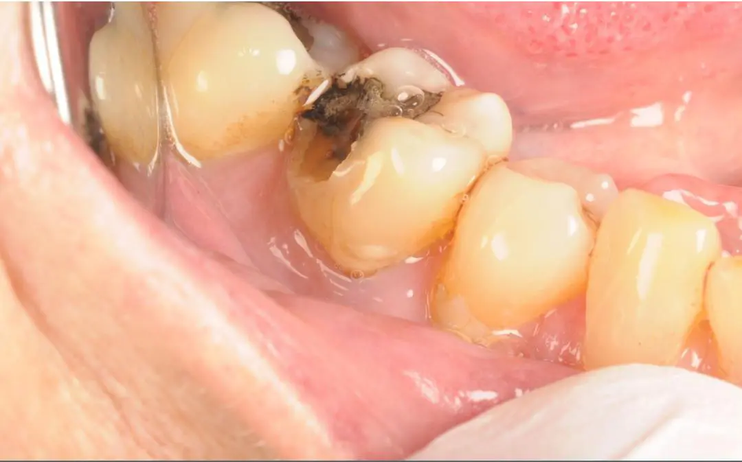
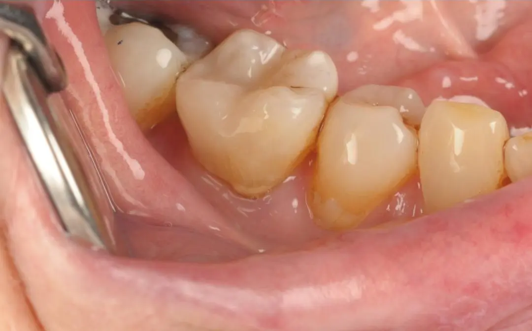
Real Patient Result: Emergency White Filling
Treatment by Dr Kamran
Even when trauma appears minor, such as a small chip or brief impact without obvious damage, radiographic assessment may still be valuable. Some injuries develop complications over time, and establishing a baseline radiographic record helps monitor any changes in tooth vitality or structure.
Children who experience dental trauma require particular attention, as injuries can affect developing permanent teeth even when only baby teeth appear damaged.
Treatment Planning Based on Radiographic Findings
Radiographic assessments provide crucial information that directly influences treatment decisions following dental trauma. The images help determine whether a tooth can be preserved, requires root canal treatment, or needs extraction and replacement.
For fractured teeth, X-rays reveal the extent and direction of fracture lines, helping dental professionals assess whether the tooth structure can support a filling, crown, or requires more extensive treatment. Root fractures visible on radiographs may necessitate splinting procedures or, in severe cases, extraction.
In cases of tooth displacement, radiographic assessment guides repositioning procedures and helps determine the appropriate stabilisation period. The images also help monitor healing progress and identify potential complications such as root resorption or infection.
Follow-up radiographic examinations are typically scheduled at regular intervals after trauma treatment to monitor healing and detect any developing complications. This ongoing assessment ensures that any changes in tooth vitality or structure are identified and addressed promptly.
Preventing Dental Trauma and Protecting Your Oral Health
While not all dental trauma can be prevented, several measures can significantly reduce the risk of injury. Using appropriate protective equipment during sports activities, particularly contact sports, provides essential protection for teeth and supporting structures.
Avoiding habits that increase trauma risk, such as using teeth as tools to open packages or chewing on hard objects like ice or pens, helps preserve tooth integrity. Teaching children about playground safety and the importance of careful behaviour during physical activities also reduces injury likelihood.
Maintaining good oral health through regular dental check-ups helps ensure that teeth remain strong and more resistant to trauma. Emergency dental services can provide immediate assessment and treatment when trauma does occur, potentially preventing complications and preserving tooth function.
Creating a dental emergency plan that includes contact information for emergency dentist services in London ensures prompt professional care when needed most.
Key Points to Remember
• Radiographic assessments are essential for identifying hidden damage following dental trauma
• Different types of dental injuries require specific imaging approaches for accurate diagnosis
• Professional evaluation should be sought promptly after any significant dental trauma
• Follow-up radiographic monitoring helps detect complications and ensures proper healing
• Preventive measures can significantly reduce the risk of dental trauma
• Early professional intervention often leads to better treatment outcomes and tooth preservation
Frequently Asked Questions
How soon after dental trauma should radiographic assessment be performed?
Radiographic assessment should ideally be performed as soon as possible after dental trauma, particularly within 24 hours of injury. Early imaging helps establish the extent of damage and guides immediate treatment decisions. Some complications may not be apparent immediately, making prompt professional evaluation crucial for optimal outcomes and long-term oral health preservation.
Are dental X-rays following trauma safe for children?
Modern digital dental X-rays use minimal radiation and are considered safe for children when clinically necessary. Following dental trauma, the benefits of radiographic assessment in identifying hidden damage and preventing complications significantly outweigh the minimal radiation exposure. Dental professionals use appropriate protective measures and take only necessary images to ensure child safety.
Can dental trauma complications develop weeks or months after the initial injury?
Yes, some dental trauma complications can develop gradually over weeks, months, or even years following the initial injury. These may include tooth discolouration, root resorption, or loss of tooth vitality. Regular radiographic monitoring helps detect these delayed complications early, allowing for appropriate intervention before more serious problems develop.
What happens if radiographic assessment reveals internal tooth damage?
When radiographic assessment identifies internal tooth damage such as root fractures or pulp injury, treatment options depend on the severity and location of the damage. Options may include monitoring with regular check-ups, root canal treatment, tooth splinting, or in severe cases, extraction and replacement. Your dental professional will discuss the most appropriate treatment based on the specific findings.
Do all types of dental trauma require radiographic assessment?
While not every minor dental trauma requires immediate radiographic assessment, most significant injuries benefit from X-ray evaluation. Even seemingly minor trauma can cause internal damage that's not visible clinically. Professional dental assessment can determine whether radiographic examination is necessary based on the injury type and clinical findings.
How often are follow-up radiographs needed after dental trauma treatment?
Follow-up radiographic schedules vary depending on the type and severity of trauma and the treatment provided. Typically, follow-up X-rays may be taken at intervals ranging from a few weeks to several months after treatment. Your dental professional will recommend an appropriate monitoring schedule based on your specific case and healing progress.
Conclusion
Radiographic assessments play a fundamental role in the proper management of dental trauma, providing essential information that guides treatment decisions and helps preserve oral health. These diagnostic tools reveal hidden damage that cannot be detected through visual examination alone, making them invaluable for accurate diagnosis and appropriate treatment planning.
Understanding the importance of prompt professional evaluation following dental trauma empowers patients to make informed decisions about their oral health care. Early radiographic assessment often leads to better treatment outcomes and can prevent the development of serious complications that might otherwise require more extensive treatment.
Maintaining good oral health through preventive measures and seeking prompt professional care when trauma occurs helps ensure the best possible outcomes for your dental health. Regular monitoring through follow-up radiographic examinations allows for early detection of any complications and appropriate intervention when necessary.
Dental symptoms and treatment options should always be assessed individually during a clinical examination.
Disclaimer
This article is for general educational purposes only and does not constitute dental advice, diagnosis, or treatment. Every patient is different, so symptoms and treatment options should be assessed by a qualified dental professional during a clinical examination. No specific outcomes are guaranteed.