
If you have a pacemaker and your dentist has suggested dental X-rays, it is entirely natural to wonder whether the procedure is safe for you. Many people with cardiac implants search online for reassurance before attending a dental appointment, wanting to understand whether dental radiography could interfere with their device or pose any risk to their health.
This article provides clear, evidence-informed guidance on dental radiography for pacemaker users, explaining the science behind how modern dental X-ray equipment works, what the current clinical evidence says about electromagnetic interference, and why open communication between your dentist and cardiologist is the most important step you can take.
Understanding your own medical history before a dental visit is a sign of good, proactive healthcare. Dental radiography remains one of the most important diagnostic tools in dentistry, and for most people — including many with pacemakers — it can be used safely with appropriate precautions and clinical assessment.
Experiencing these symptoms?
Delaying treatment can lead to tooth loss. We have slots available today.
Is Dental Radiography Safe for Pacemaker Users?
Current clinical evidence suggests that modern dental X-ray equipment, which operates at low radiation doses and very short exposure times, poses minimal risk of electromagnetic interference with contemporary pacemakers. However, dental radiography for pacemaker users should always be preceded by a thorough medical history review and, where appropriate, consultation with the patient's cardiologist.
Understanding Pacemakers and Why Patients Have Concerns
A pacemaker is a small implanted electronic device designed to regulate abnormal heart rhythms. It delivers tiny electrical impulses to the heart muscle when it detects an irregular beat or a pause in the heart's natural rhythm. Given that pacemakers are sensitive electronic devices, it is reasonable for patients and clinicians to consider whether external electromagnetic energy sources — including those used in dentistry — could affect their function.
Historically, concerns arose because older pacemaker models were more susceptible to electromagnetic interference (EMI) from certain medical and dental equipment. These concerns, although largely resolved with advances in technology, have persisted in public awareness, leading many patients to feel uncertain about routine dental procedures.
Meet Dr. Yasha Shirazi
Principal Dentist at Emergency Dentist London
"We treat hundreds of dental emergencies every month. The sooner you come in, the easier the fix usually is."
Book an appointment with our team →Today, pacemakers manufactured over the past two decades are significantly better shielded against EMI. Most modern devices are designed to be resistant to the low-level electromagnetic fields generated by standard dental equipment. Nevertheless, clinical caution and patient-specific assessment remain the professional standard of care.
It is important to note that every patient's situation is individual. The type of pacemaker, the reason it was implanted, and any additional cardiac conditions all play a role in how your dental team approaches your care.
How Dental X-Rays Work: A Clinical Explanation
Dental radiography involves the use of ionising radiation to produce diagnostic images of the teeth, jaw, and surrounding structures. Modern dental X-ray equipment uses carefully controlled, low-dose radiation beams directed at a specific area of the mouth for a very brief exposure time — typically less than one second for a standard periapical or bitewing radiograph.
The electromagnetic spectrum used in dental X-rays is fundamentally different from the radio-frequency electromagnetic fields that older pacemakers were most susceptible to. Dental X-ray machines do not generate significant radio-frequency (RF) emissions during normal operation. The radiation produced is ionising in nature but is highly localised, directional, and does not meaningfully interact with the electronic components of an implanted cardiac device.
Additionally, modern dental practices use digital radiography systems, which require even lower radiation doses than traditional film-based X-rays. The combination of precise beam collimation, short exposure times, and digital sensor technology means that the overall radiation output — and any theoretical electromagnetic field generated — is minimal.
Understanding this distinction between ionising radiation and radio-frequency electromagnetic fields helps clarify why dental X-rays are generally considered low-risk for pacemaker users, though individual clinical assessment is always essential.
For a broader overview of advanced imaging in urgent and complex cases, see our CBCT and 3D imaging clinical guide.
Current Clinical Evidence and Professional Guidance
The weight of clinical evidence available to date suggests that contemporary dental radiography equipment does not produce levels of electromagnetic interference sufficient to affect modern pacemakers in a clinically significant way.
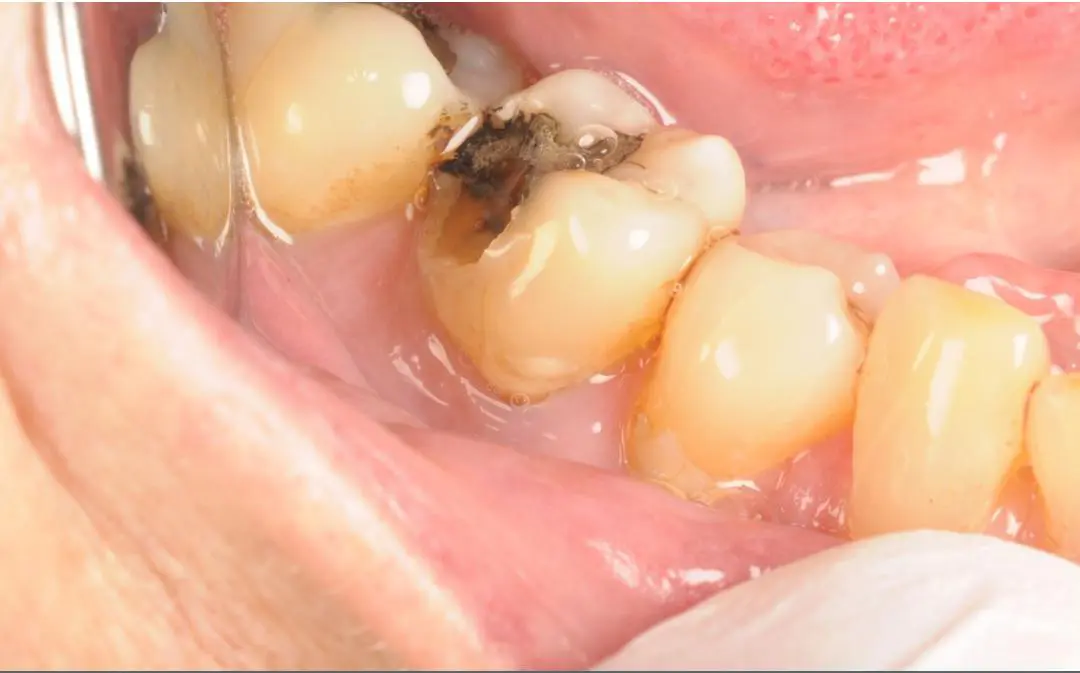
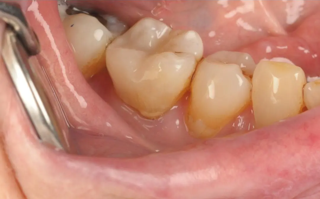
Real Patient Result: Emergency White Filling
Treatment by Dr Kamran
Several key bodies and research findings inform current practice:
- The British Dental Association (BDA) and the British Heart Rhythm Society both acknowledge that dental X-ray equipment operates at exposure levels and frequencies that are unlikely to interfere with modern implantable cardiac devices.
- Research published in peer-reviewed dental and cardiology journals consistently indicates that the risk of clinically meaningful EMI from dental radiography is very low with current-generation pacemakers.
- The General Dental Council (GDC) requires that dentists take a comprehensive medical history before treatment, which includes documenting the presence of any cardiac implantable electronic devices (CIEDs).
It is important to emphasise that clinical guidance is not a substitute for individual assessment. Older pacemaker models, rare device configurations, or patients with specific cardiac vulnerabilities may require additional precautions. This is why disclosing your pacemaker to your dental team — and updating them if your device is replaced or reprogrammed — is so important.
Pre-Appointment Communication: What Patients Should Do
One of the most effective steps a pacemaker user can take before any dental appointment is to ensure their dental team is fully informed about their cardiac implant. This may seem straightforward, but many patients assume their dentist already knows, or they may be uncertain which details are relevant.
Here is what patients with pacemakers are generally advised to do before dental radiography:
Inform your dentist at the earliest opportunity. When booking or attending your appointment, let reception and your dentist know that you have a pacemaker. This should be documented clearly in your medical history.
Provide device details if possible. If you have a pacemaker identification card (which cardiac device patients are typically given), bring it with you. This card contains the manufacturer name, model number, and implant date, which can help your dentist and, if needed, your cardiologist assess any relevant considerations.
Speak to your cardiologist if you have specific concerns. For patients with complex cardiac histories or older pacemaker models, your dentist may recommend a brief consultation with your cardiologist or the cardiac device clinic before proceeding with radiography.
Do not avoid necessary dental X-rays without clinical advice. Dental radiography is an important diagnostic tool. Avoiding it without professional guidance could mean that dental problems go undetected, potentially leading to more complex issues later.
You can learn more about how our team approaches patients with complex medical histories by visiting our emergency dental assessment page.
The Role of the Dental Team in Safe Radiographic Practice
Your dental team has professional and regulatory responsibilities when it comes to radiographic safety. Under the Ionising Radiations Regulations 2017 (IRR17) and the Ionising Radiation (Medical Exposure) Regulations 2017 (IR(ME)R), dental practices in the UK are required to justify every radiographic exposure and ensure that radiation doses remain as low as reasonably practicable (the ALARP principle).
For pacemaker users, this professional framework provides an additional layer of protection, as your dentist should:
- Record your medical history accurately, including the presence of any cardiac implantable electronic devices
- Consider whether radiography is clinically justified for your specific diagnostic needs
- Select the appropriate radiographic technique — for example, choosing periapical or bitewing X-rays over panoramic radiography where clinically suitable, if there are any specific concerns about the patient's device
- Liaise with medical colleagues when the patient's history warrants further advice before proceeding
Dental nurses and radiographers working within the practice also play an important role in ensuring correct technique, appropriate shielding, and accurate patient history documentation.
This collaborative approach ensures that every patient — including those with cardiac implants — receives safe, responsible, and clinically appropriate care.
Types of Dental Radiography and Considerations for Pacemaker Users
There are several different types of dental X-ray that patients may encounter, and it is helpful to understand what each involves:
Periapical radiographs show the whole tooth, from crown to root tip, and the surrounding bone. They use a very small, directed beam and brief exposure time. These are among the most commonly used dental X-rays and are generally considered to present no meaningful electromagnetic concern for pacemaker users.
Bitewing radiographs show the upper and lower back teeth in a single image and are commonly used to detect decay between teeth. Like periapical X-rays, the exposure is brief and localised.
Panoramic radiographs (OPG or DPT) provide a broad view of the entire dentition, jaw, and surrounding structures. The X-ray source rotates around the patient's head. This type involves slightly longer exposure than intraoral X-rays, though radiation doses remain within safe clinical limits. There is no strong evidence that panoramic radiography presents a significantly elevated EMI risk for modern pacemakers, but dentists should note the device type and apply clinical judgement.
Cone Beam Computed Tomography (CBCT) is a more advanced three-dimensional imaging technique used in specific clinical situations such as implant planning. CBCT involves higher radiation dose than standard dental X-rays and should only be used when clinically justified. For patients with pacemakers, CBCT should be discussed carefully with the dental team.
For patients requiring more complex treatment planning, our guide to dental implants and crowns for long-term restorative care explains how advanced imaging is used responsibly.
When Professional Dental Assessment May Be Needed
If you have a pacemaker and are experiencing dental symptoms, it is important not to delay seeking professional assessment. Dental problems left untreated can escalate, and some oral infections can have broader health implications — something particularly relevant for patients with cardiac conditions.
Consider seeking prompt dental advice if you notice any of the following:
- Persistent toothache or jaw pain that does not resolve on its own
- Swelling around the face, jaw, or neck — this can sometimes indicate a spreading dental infection, and these warning signs of tooth infection spread are useful to recognise early
- Sensitivity to hot, cold, or sweet foods and drinks that appears suddenly or worsens over time
- Bleeding gums, particularly if persistent or unexplained
- A tooth that feels loose, especially in an adult
- Difficulty opening the mouth or swallowing, which should be assessed promptly
None of these symptoms are causes for alarm, but they are signs that a professional dental assessment would be appropriate. Your dentist can evaluate what is happening and advise on the most suitable course of action, including whether radiography is needed to assist diagnosis.
Watch the video below for information about dental emergency consultations in London, including costs and membership options:
Oral Health Maintenance for Patients with Cardiac Implants
Maintaining good oral health is important for everyone, and there is well-established evidence linking oral health to overall systemic wellbeing. For patients with cardiac conditions, including those with pacemakers, maintaining healthy teeth and gums is a particularly meaningful aspect of general health management.
Here are some practical recommendations for maintaining oral health as a pacemaker user:
Attend regular dental check-ups. Routine dental examinations allow problems to be identified early, reducing the likelihood of needing more extensive treatment. If you have concerns about dental procedures and your pacemaker, discussing these at a routine appointment — rather than waiting until a problem arises — is always a sensible approach.
Maintain a consistent oral hygiene routine. Brushing twice daily with fluoride toothpaste and cleaning between teeth daily with floss or interdental brushes helps to prevent tooth decay and gum disease.
Stay hydrated and maintain a balanced diet. Reducing sugary foods and drinks protects tooth enamel and reduces the risk of decay. Staying well hydrated supports saliva production, which has a natural protective role in the mouth.
Inform all healthcare providers about your full medical history. Your dentist, cardiologist, and GP should all have accurate and up-to-date information about your medications and implanted devices, as some dental medications and treatments can interact with cardiac medications.
Our preventative oral care guide provides further guidance on maintaining excellent oral health through routine care.
Key Points to Remember
- Dental radiography for pacemaker users is generally considered safe with modern equipment, though individual clinical assessment is always required before proceeding.
- Modern pacemakers are significantly better shielded against electromagnetic interference than older models, and dental X-ray equipment operates at frequencies and exposure levels unlikely to cause clinically significant interference.
- Always inform your dental team that you have a pacemaker — and provide device details if you have them — before any X-rays are taken.
- Your dentist has a professional and regulatory obligation to take a thorough medical history and justify every radiographic exposure.
- If you have concerns about your specific pacemaker model, your dentist may recommend consultation with your cardiologist before radiography.
- Maintaining regular dental check-ups and a good daily oral hygiene routine is particularly important for patients with cardiac conditions.
Frequently Asked Questions
Can dental X-rays affect my pacemaker?
Current clinical evidence suggests that modern dental X-ray equipment is very unlikely to interfere with contemporary pacemakers in a clinically meaningful way. Dental X-rays operate using ionising radiation with short, localised exposures, rather than the radio-frequency electromagnetic fields that older pacemaker models were most sensitive to. However, every patient's situation is individual, and your dentist will review your medical history carefully before proceeding with any radiography. If you have an older pacemaker model or specific cardiac concerns, your dentist may recommend speaking with your cardiologist beforehand.
Do I need to tell my dentist I have a pacemaker before dental X-rays?
Yes — it is very important to inform your dental team that you have a pacemaker, ideally before your appointment. Your dentist is required to record your full medical history before carrying out any dental procedure, including radiography. Disclosing your pacemaker allows the dental team to apply appropriate clinical judgement, select the most suitable radiographic technique, and, where necessary, liaise with your cardiologist. Bringing your pacemaker identification card to the appointment can also be helpful, as it provides the device model and manufacturer details.
Are there any types of dental X-ray that are safer for pacemaker users?
Standard intraoral X-rays — such as periapical and bitewing radiographs — involve very short exposures and highly localised beams, and are generally considered appropriate for pacemaker users following a medical history review. Panoramic X-rays (OPGs) have slightly longer exposures but remain within safe limits, with no strong evidence of elevated EMI risk for modern devices. Cone Beam CT (CBCT) involves higher radiation doses and should only be used when clinically justified. Your dentist will recommend the most appropriate type based on your clinical needs and device history.
What should I bring to my dental appointment if I have a pacemaker?
It is helpful to bring your pacemaker identification card, which is usually issued at the time of device implantation and contains details such as the device manufacturer, model number, and implant date. You should also be prepared to provide information about any cardiac medications you are taking, as some medications can be relevant to dental treatment planning. If you have recently had your pacemaker replaced, upgraded, or reprogrammed, inform your dentist of this at your appointment, as device specifications may have changed.
Could a dental infection be more serious for someone with a pacemaker?
Dental infections should always be assessed and treated promptly, and this is particularly relevant for patients with cardiac implants. There is an established body of evidence linking untreated oral infections and advanced gum disease to systemic health outcomes, including cardiovascular implications. Bacteria from oral infections can, in rare circumstances, enter the bloodstream. If you have a pacemaker and notice signs of a dental infection — such as persistent pain, swelling, or a bad taste in the mouth — seeking professional dental assessment promptly is advisable. Your dental team will assess the situation and advise on appropriate treatment.
Will my dentist refuse to take X-rays because I have a pacemaker?
It is unlikely that a dentist would refuse necessary X-rays solely because a patient has a pacemaker, as current evidence supports the safety of modern dental radiography for most pacemaker users. However, your dentist may take additional steps, such as reviewing your device details, consulting with your cardiologist, or selecting a particular radiographic technique, before proceeding. If you have concerns, speaking openly with your dentist about your pacemaker and any questions you have is the best approach. Good communication between patient and clinician is central to safe, patient-centred dental care.
Conclusion
Dental radiography for pacemaker users is a topic that understandably raises questions, but the current clinical evidence and professional guidance provides a reassuring picture for most patients. Modern dental X-ray equipment operates at exposure levels that are very unlikely to interfere with contemporary pacemaker devices, and the dental profession has robust regulatory frameworks in place to ensure every patient receives safe, justified radiographic care.
The most important steps any pacemaker user can take are to keep their dental team fully informed about their cardiac implant, attend regular dental check-ups, and not delay seeking advice when dental symptoms arise. Maintaining good oral health is not just about your teeth — it is an important part of your overall wellbeing, particularly when you are managing a cardiac condition.
If you have concerns about dental X-rays and your pacemaker, speaking with your dentist is always the right first step. They can review your individual circumstances, consider your device history, and — where appropriate — liaise with your cardiologist to ensure your care is as safe and comfortable as possible.
Dental symptoms and treatment options should always be assessed individually during a clinical examination.
Disclaimer
This article is for general educational information only and does not constitute dental or medical advice.
Diagnosis and treatment suitability can only be confirmed by a qualified clinician during an in-person examination.
Do not use this content as a substitute for personalised professional advice based on your medical history.
No treatment outcomes are guaranteed, and decisions should be made with your treating dental and medical team.