
If you have been referred for an MRI scan and have metal dental crowns, it is entirely natural to have questions about whether it is safe to proceed. Many patients search online after receiving an MRI referral, unsure of what their dental restorations might mean for the procedure. The concern is understandable — after all, MRI machines use powerful magnetic fields, and the thought of having metal in your mouth during such a scan can feel daunting.
This article aims to address those concerns clearly and responsibly. MRI scans in patients with metal dental crowns is a topic that spans both medical and dental care, and understanding the considerations involved can help you have more informed conversations with your dentist, radiologist, or hospital team.
We will explore how different types of metal crowns interact with MRI technology, what questions to raise with your clinical team, and when professional dental assessment may be appropriate. This information is educational and does not replace individual clinical advice.
Experiencing these symptoms?
Delaying treatment can lead to tooth loss. We have slots available today.
Are Metal Dental Crowns Safe During an MRI Scan?
In most cases, patients with metal dental crowns can safely undergo MRI scans, as dental restorations are generally securely fixed within the mouth. However, the type of metal used in the crown and its proximity to the area being scanned can affect image quality. Patients should always inform their MRI radiographer and dental team before proceeding.
Understanding Metal Dental Crowns: A Brief Background
Dental crowns are restorations placed over damaged, weakened, or significantly decayed teeth to restore their function and appearance. They are one of the most commonly used restorations in both NHS and private dentistry.
Metal crowns have been used in dentistry for many decades and remain a durable option, particularly for back teeth where chewing forces are greatest. These restorations may be made from a variety of metallic alloys, including:
Meet Dr. Yasha Shirazi
Principal Dentist at Emergency Dentist London
"We treat hundreds of dental emergencies every month. The sooner you come in, the easier the fix usually is."
Book an appointment with our team →- Gold alloys — historically one of the most biocompatible and durable crown materials
- Base metal alloys — often containing nickel, chromium, or cobalt
- Porcelain-fused-to-metal (PFM) crowns — which combine a metal substructure with a porcelain outer layer
More recently, tooth-coloured materials such as zirconia and all-ceramic crowns have become increasingly popular alternatives, partly due to aesthetic preferences. However, many patients in the UK still have older metal or PFM crowns in place, which is why questions around MRI compatibility remain relevant and worth understanding.
If you are considering updating your existing restorations, reviewing clinical guidance for managing a dislodged or loose dental crown may be a helpful starting point before discussing material suitability with your dentist.
How MRI Scanners Work and Why Metal Is a Consideration
MRI (Magnetic Resonance Imaging) scanners use powerful magnetic fields and radiofrequency pulses to produce detailed images of soft tissues, organs, and structures within the body. Unlike X-rays, MRI does not use ionising radiation, which is one reason it is frequently used for neurological, musculoskeletal, and soft tissue imaging.
The concern surrounding metal and MRI scans broadly falls into two categories:
1. Safety Risks from Ferromagnetic Materials
Strongly ferromagnetic metals — those that are significantly attracted to magnets — can pose genuine safety risks inside an MRI scanner. These materials may be subject to movement, heating, or interference with the magnetic field. However, most dental alloys used in crown fabrication are either non-ferromagnetic or only weakly magnetic, meaning they are unlikely to create safety hazards under standard MRI conditions.
2. Image Artefact and Diagnostic Interference
Even when metal dental restorations do not pose a direct safety risk, they can cause what is known as magnetic susceptibility artefact — a distortion or signal void in the MRI image around the area of the metal. This is particularly relevant when the region being imaged is in close proximity to the mouth, jaw, or skull base.
For scans of areas such as the abdomen, spine, or limbs, the presence of dental crowns is unlikely to have any significant impact on image quality or clinical utility.
The Clinical Science: How Different Crown Materials Behave in an MRI Environment
Understanding the underlying material science helps explain why some crowns raise greater concerns than others during MRI imaging.
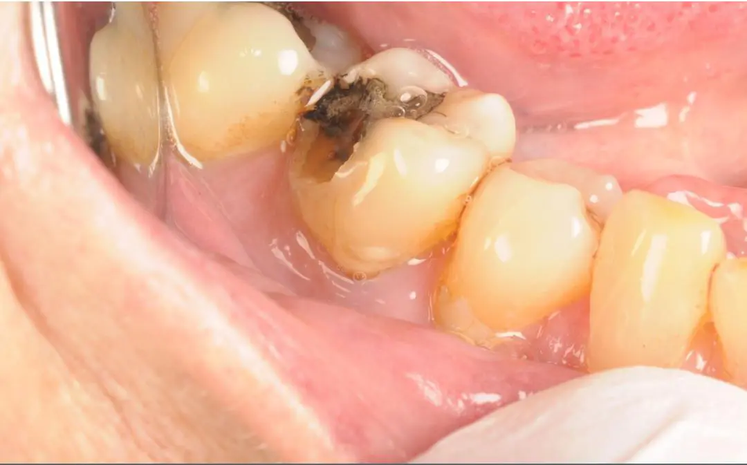
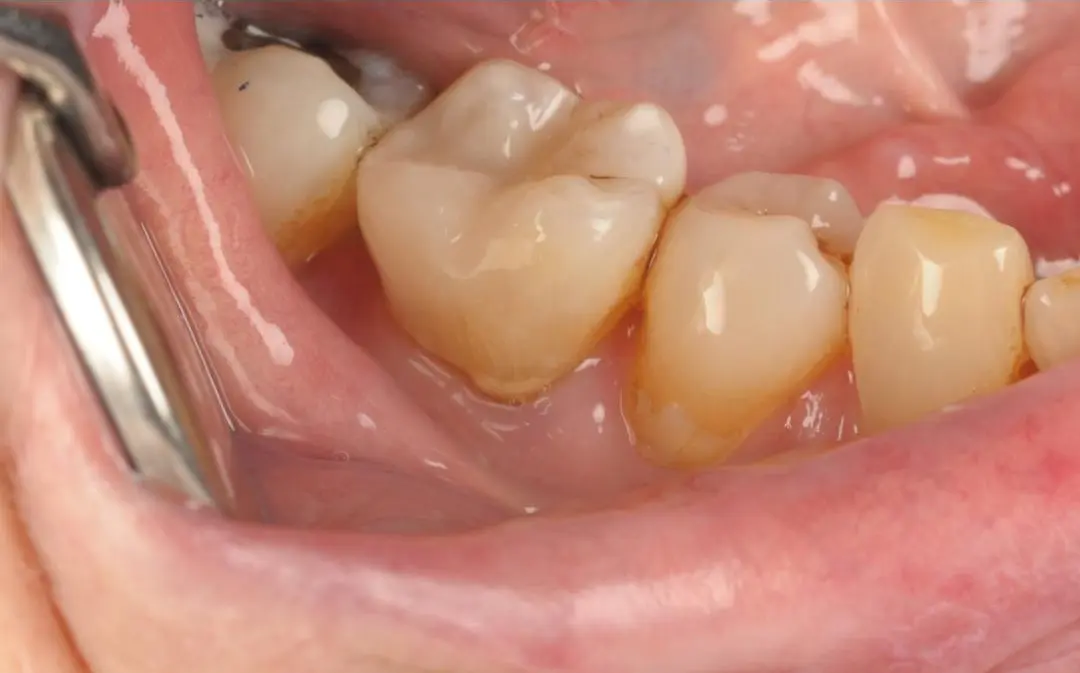
Real Patient Result: Emergency White Filling
Treatment by Dr Kamran
Gold alloys are generally non-ferromagnetic and have been widely studied in relation to MRI safety. They tend to have a low magnetic susceptibility, meaning they cause minimal disruption to the magnetic field and produce relatively little artefact.
Base metal alloys, particularly those containing cobalt-chromium or nickel-chromium, can have variable magnetic properties depending on their precise composition. Some may cause more notable image distortion, particularly in head and neck imaging.
Porcelain-fused-to-metal (PFM) crowns depend on the specific metal substructure used. The porcelain component itself is inert and does not interact with the magnetic field, but the metal beneath can still produce artefact.
Zirconia and all-ceramic crowns are essentially inert in an MRI environment and do not cause ferromagnetic interaction or significant image distortion.
It is important to note that even crowns which cause artefact do not typically pose a physical safety risk to the patient — the concern is primarily one of imaging quality. Whether that artefact is clinically significant depends entirely on the anatomical region being examined.
Communicating with Your Medical and Dental Teams Before an MRI
Clear communication between a patient, their dentist, and the referring medical team is an essential part of safe MRI preparation.
Before attending for your MRI scan, it may be helpful to:
- Inform the MRI radiographer that you have dental crowns and, if possible, advise what material they are made from
- Contact your dentist to request documentation about the type and composition of your existing crowns where records are available
- Ask the radiographer or radiologist whether your specific dental restorations are likely to affect the quality of the images for the anatomical region being scanned
Hospital MRI units typically carry out a safety screening process prior to all scans. Patients are routinely asked about the presence of metallic implants, restorations, and devices. Dental crowns should always be declared at this stage — even if they seem unlikely to be relevant.
In some cases, the radiologist may choose to adjust scan parameters or note the likely artefact in their report. In other cases, they may determine that the crowns are not a concern for the region in question.
When Professional Dental Assessment May Be Appropriate
There are several situations where seeking dental advice before or after an MRI scan may be beneficial.
If you are unsure what material your existing crowns are made from, your dentist may be able to review your dental records or take a clinical look at your restorations. Older crowns may not always have clear documentation, particularly if they were placed many years ago or by a different provider.
Additionally, if you have been experiencing any of the following alongside your forthcoming scan, it would be sensible to arrange a dental review:
- Discomfort or sensitivity around a crowned tooth that has persisted for more than a few days
- A crown that feels loose, cracked, or structurally compromised
- Swelling or tenderness around the gums near a crowned tooth
- A noticeable change in your bite since a crown was placed
None of these symptoms are automatically cause for alarm, but each is a valid reason to seek professional assessment. A dentist can evaluate the condition of your existing restorations and advise on whether any action is appropriate before or after your MRI, including whether lost crown repair assessment may be relevant.
Emergency Dental Support: When You Need Advice Quickly
If you are due to have an MRI scan soon and have a dental concern you would like reviewed promptly, accessing timely dental support is important. Our team in London may offer same-day appointments for patients who need urgent dental assessment, subject to availability.
Whether you have concerns about an existing crown, are experiencing unexpected dental discomfort, or need a clinical review ahead of a medical procedure, an emergency dental appointment in London can help ensure you have the information you need before proceeding with other investigations.
Impact on MRI Image Quality: What Patients Should Realistically Expect
For most patients with metal dental crowns, the practical impact on an MRI scan depends largely on the area of the body being examined.
When the scan is distant from the mouth (for example, the knee, hip, lumbar spine, or abdomen), dental metal restorations are unlikely to cause any meaningful interference with image quality. Radiologists are experienced in interpreting scans in patients with various dental restorations, and the presence of crowns rarely prevents useful diagnostic imaging in these regions.
When the scan is of the head, face, neck, or skull base, the situation requires more careful consideration. In these cases, metallic artefact from dental crowns can obscure nearby anatomical structures, making it more difficult to interpret certain areas of the scan. Radiologists will be aware of this and will take it into account when reporting findings.
There are specific imaging techniques, including adjusted pulse sequences and positioning approaches, that can help to reduce the extent of dental artefact in head and neck MRI. These decisions are made by the radiologist and MRI technologist based on the clinical priorities of the scan.
It is worth noting that the presence of dental crowns does not mean an MRI cannot proceed — it simply means that the clinical team will account for the restorations as part of their overall assessment.
Considering Crown Material Options: A Note on Modern Alternatives
For patients who are aware that they may require regular MRI scanning in the future — for example, due to a chronic health condition or neurological monitoring — it may be worth discussing crown material preferences with their dentist proactively.
Tooth-coloured materials such as zirconia and all-ceramic crowns produce no ferromagnetic interaction and minimal or no image artefact. For patients requiring crowns on visible front teeth or those with known imaging requirements, these materials offer both aesthetic benefits and MRI compatibility.
That said, material choice should always be guided by clinical suitability for each individual tooth and patient — not by MRI considerations alone. Factors including the position of the tooth, the level of biting force it endures, the extent of the preparation, and the patient's overall oral health all inform which material is most appropriate.
Discussing tooth bonding vs dental crowns with a qualified dentist can help you understand your options based on your specific clinical circumstances.
Prevention and Oral Health Advice for Patients with Dental Crowns
Maintaining good oral health around dental crowns can help prolong their lifespan and reduce the likelihood of complications. Here are some practical tips:
- Brush twice daily with fluoride toothpaste, paying careful attention to the gum line around each crown
- Floss daily to remove plaque from between crowned teeth, as the junction between the crown and the gum can be susceptible to plaque accumulation
- Attend regular dental check-ups — your dentist can monitor the condition of your crowns and identify early signs of wear or cement failure; if you need to arrange one promptly, book a dental appointment
- Avoid biting on very hard foods or objects that could fracture or dislodge a crown
- Report any changes promptly — if a crown feels loose, different in bite, or if the surrounding gum looks inflamed, arrange a dental review
- Inform all healthcare providers about your dental restorations when relevant, including before medical imaging procedures
Crowns are durable restorations, but like all dental work, they benefit from attentive maintenance and regular professional review.
Key Points to Remember
- MRI scans in patients with metal dental crowns are in most cases safe to proceed, but clinical assessment of crown type and scan region is important
- The primary concern with metal crowns in MRI is image artefact (distortion) rather than direct physical harm, in most cases involving typical dental alloys
- Always declare your dental restorations to the MRI radiographer during the standard safety screening process
- The impact of dental crowns on MRI image quality depends on the type of metal, the specific alloy composition, and which area of the body is being scanned
- Gold and ceramic-based crowns generally have a lower impact on MRI imaging than some base metal alloys
- If you have concerns about your existing crowns before an MRI, a dental review can help clarify what materials are present and support informed decision-making
Frequently Asked Questions
Is it safe to have an MRI scan if I have a metal dental crown?
In the majority of cases, patients with metal dental crowns can safely undergo MRI scanning. Most dental alloys used in crown fabrication have low or minimal ferromagnetic properties, meaning they are unlikely to pose a direct physical safety risk during the scan. However, it is important to inform the radiographer about all dental restorations before the procedure so that appropriate precautions can be taken. The safety team at the MRI unit will assess your specific situation as part of their standard screening process.
Will my metal crown affect the quality of my MRI images?
This depends on where in the body the scan is being performed. If the MRI is focused on an area far from your mouth — such as the spine, abdomen, or limbs — dental crowns are unlikely to have a meaningful effect on image quality. For head and neck MRI scans, metallic dental restorations may cause image artefact (distortion) in nearby areas. Radiologists are trained to account for this and will document any relevant limitations in their report.
Do I need to tell anyone about my dental crowns before an MRI?
Yes. You should always declare your dental crowns — and any other dental restorations — during the MRI safety screening questionnaire. This allows the radiography team to assess whether the materials involved may affect the scan or the images. If possible, it can be useful to contact your dentist beforehand to find out what type of metal was used in your crown, particularly if it was placed some years ago.
Are tooth-coloured crowns better than metal crowns for patients who need regular MRI scans?
Tooth-coloured restorations such as zirconia and all-ceramic crowns are generally inert in MRI environments and do not cause ferromagnetic interaction or significant image artefact. For patients who require regular MRI monitoring for an ongoing health condition, this may be worth discussing with their dentist when considering future dental restorations. However, crown material selection should always be based on clinical suitability first, taking into account the location of the tooth, biting forces, and the individual patient's oral health needs.
What should I do if my dental crown feels uncomfortable before or after my MRI?
If you notice any discomfort, sensitivity, looseness, or change around a crowned tooth — whether related to your MRI scan or not — it is advisable to arrange a dental review. While an MRI scan itself would not physically damage a properly fitted, non-ferromagnetic dental crown, any pre-existing issues with a restoration may merit attention. A dentist can examine the tooth and crown, assess the surrounding gum tissue, and advise on whether any treatment or monitoring is needed.
Can an MRI scan heat up or move a metal dental crown?
Properly fitted dental crowns made from typical dental alloys are generally not at significant risk of movement or clinically meaningful heating during a standard MRI scan. Unlike certain implanted medical devices, dental crowns are cemented restorations with minimal mass and are typically not made from strongly ferromagnetic materials. That said, any concerns should always be discussed with the radiography team before the scan, as they will be best placed to assess the specific materials involved and the parameters of the MRI being performed.
Conclusion
Understanding the clinical considerations for MRI scans in patients with metal crowns is an important part of being an informed patient. For most people, the presence of dental crowns does not prevent MRI imaging from taking place safely. The key lies in clear communication between you, your dental team, and your medical team — ensuring that the right information is shared before the scan so that everyone involved can make appropriate decisions.
Whether the concern relates to safety, image quality, or simply uncertainty about what materials your existing crowns contain, professional dental advice can help provide clarity. If you have questions about your crowns ahead of an upcoming scan, a dental assessment is always a sensible step.
Dental symptoms and treatment options should always be assessed individually during a clinical examination.
If you are experiencing any dental concerns or would like advice about your existing restorations, we encourage you to seek professional dental guidance from a qualified dentist who can review your individual circumstances.
Disclaimer
This article is for general educational information only and does not constitute dental or medical advice.
It does not replace a face-to-face assessment by a GDC-registered clinician.
Treatment suitability and outcomes vary by individual and can only be confirmed after clinical examination.
This content is prepared in line with GDC, CQC, and ASA CAP communication principles.